Pinokioputhzy
Member
end of march this year my horse was lame, vet came in April. This was her diagnosis:
General - Anamnesis: Lianne (owner) is abroad for an internship; her father is present.
The rider noticed that Pino did not move entirely regularly during riding and doubted whether he was lame. No swellings or heat were felt. Known history of a previous injury to the SDFT (Superficial Digital Flexor Tendon) in the right front leg.
Findings:
Inspection/Palpation:
Examination:
Ultrasound of the fetlock joint and suspensory ligaments:
12-04-2024:
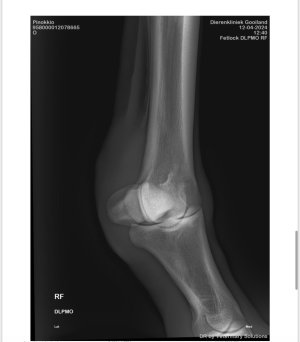
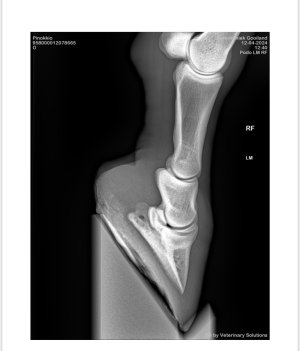
X-rays of the fetlock joint:
Chronic strain on the suspensory apparatus due to the position of the feet and an imbalance in the hoof-pastern axis, increasing the strain on the tendons and ligaments.
Treatment/Advice:
At this moment we are on our old dressage level, which we did before the injury.
But what about jumping? My vet was not clear about this. Ofcourse I will call them also

General - Anamnesis: Lianne (owner) is abroad for an internship; her father is present.
The rider noticed that Pino did not move entirely regularly during riding and doubted whether he was lame. No swellings or heat were felt. Known history of a previous injury to the SDFT (Superficial Digital Flexor Tendon) in the right front leg.
Findings:
Inspection/Palpation:
- No heat, swelling, or fluid buildup.
- Dry legs, hoof joints symmetrical with flat soles, slightly underrun heels, and a slightly dropped fetlock.
Examination:
- Straight hard surface (walk): No abnormalities.
- Straight hard surface (trot): 1/10 lameness in the right front leg (RF).
- Lunging on hard surface: 1/5 irregularity in both trot and walk on the RF.
- Lunging on soft surface: 1/10 irregularity in trot, none in walk.
- Flexion test: Slightly positive distally in the left front leg (LF).
Ultrasound of the fetlock joint and suspensory ligaments:
- Old lesion still visible in the deep digital flexor tendon (calm and stable, no changes).
- No inflammation in the fetlock joint.
- Mild chronic swelling of the medial interosseous ligament, slightly more pronounced on the RF compared to the LF.
- Medial collateral ligament slightly thickened.
- Medial collateral ligament of the coffin joint slightly enlarged laterally.
12-04-2024:
X-rays of the fetlock joint:
- Mild sclerosis and a small area of lucency in the medial sesamoid bones due to chronic strain on the suspensory apparatus.
- Minimal negative palmar angle in the RF.
- Sole still thin and not well-rounded.
Chronic strain on the suspensory apparatus due to the position of the feet and an imbalance in the hoof-pastern axis, increasing the strain on the tendons and ligaments.
Treatment/Advice:
- Corrective shoeing:
- Shorten the toes.
- Use tendon-supporting shoes with a wide toe and a slightly raised sole for better shock absorption.
- 4–6 weeks of hand walking, max. 20 minutes per day.
- After 4–6 weeks, clinical re-evaluation to assess progress and determine how to gradually increase activity.
At this moment we are on our old dressage level, which we did before the injury.
But what about jumping? My vet was not clear about this. Ofcourse I will call them also


 )
)