Dave's Mam
Well-Known Member
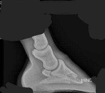
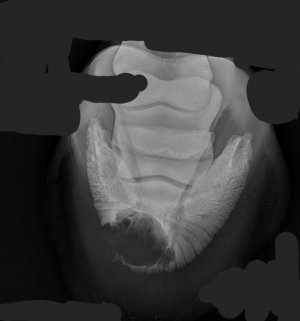
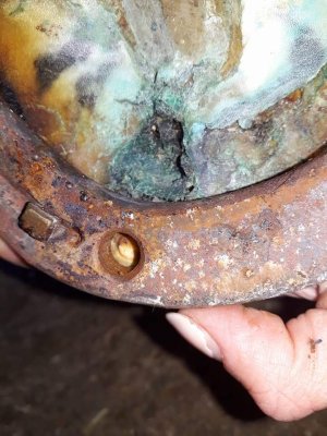
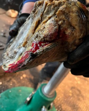
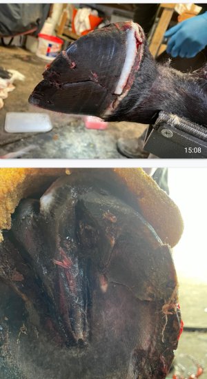
Canker?Shall we go to feet? I have lots of different kinds of feet pics!
Oooh! Could make some money on Only Fans for those with foot fettish!
Canker?Shall we go to feet? I have lots of different kinds of feet pics!
Oooh! Could make some money on Only Fans for those with foot fettish!
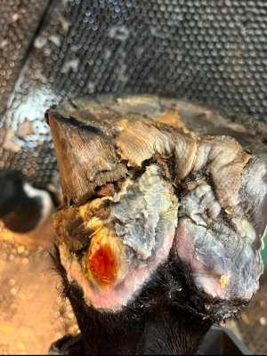
 some more obvious than others.
Hmmm keratoma?
I guessed hole in a tendon 'somewhere'?Close but no Cigar. The big black hole is a tear in a Superficial Digital Flexor Tendon. It's the big one you feel running down the back of your horses cannon bone.
This was too much of a hole and also a reinjury of the same area to make stem treatment possible. So box rest, walking then turned away for a few months. The horse did race again.
That is a very close up. I'll see if I can find my wider one with all layers in.
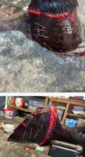
That is a whole load of staples!!!Luckily there was no damage to the underlying structures. But the racecourse vet was a bit staple happy!
EYP?I’m liking this game… will try to dig out some horse ones (they aren’t as good as yours) but in the meantime please enjoy these bird ones (why did we have to PTS?)
1
View attachment 118574
2
View attachment 118577
That is a curious angle - is it showing part of it magnified?Shall we go to feet? I have lots of different kinds of feet pics!
Oooh! Could make some money on Only Fans for those with foot fettish!
Gutted I missed the condylar fracture one, I was ready with my lag screw fixation as the answer!
Anyone who is interested in cool fracture repairs, I highly recommend the exceptional Dr Patty Hogan who is a NY based orthopaedic specialist.
Gelding according to the post.They have made the fetlock rigid, how can that ever be "sound" ?
Sound enough to sell its sperm, maybe!
They have made the fetlock rigid, how can that ever be "sound" ?
Sound enough to sell its sperm, maybe!
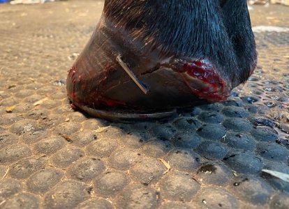
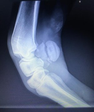
See if you can spot the problem and guess how lame he was......will post the MRI and follow scans after
Pedal bone fracture? Probably 1/5 lame?


Not quite, would have ended up that way, thats the other view, sound apart from a few odd strides and bucking going into canter on the right rein, its the rh hoof
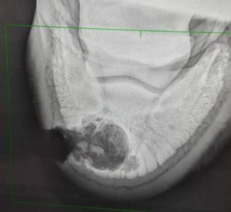
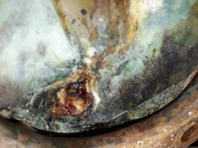
Keratoma?
Yep, lady to reported thw MRI results said it was one of the biggest she has ever seen.
He was back in walk work 4 months after and a year later back in full work
MRI
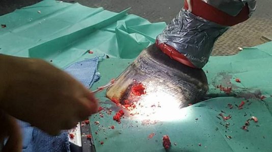
Picture mid op (2017)
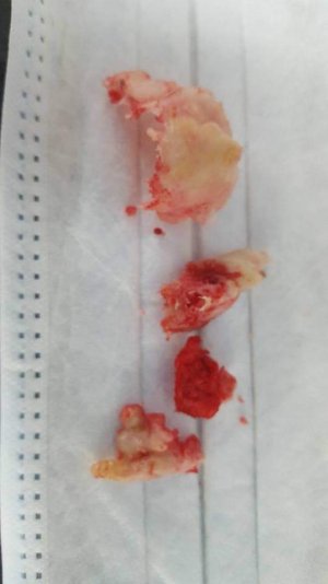
The keratoma
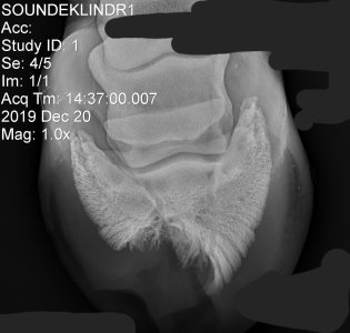
And follow up xray 2019
Number 1I'll leave you with a few more feet to muse over whilst I go to bed
I have more pics of some of these from the initial problem to fixing it.
That is a CT scan of an Atlanto-occipital joint showing an exostosis AKA a small bony growth of (probably of the first cervical vertebra of the neck) which does not impinge on the spinal cord.View attachment 118586View attachment 118587
I don’t have undoctored images of this but this is the most random equine diagnostic image I’ve got I think. Anyone like to guess the imaging modality and what the arrow is pointing at?
That is seriously impressive! Just out of interest, how long did it take for the hoof to regrow?
Yep here’s the 3d reconstructionNumber 1
That is a CT scan of an Atlanto-occipital joint showing an exostosis AKA a small bony growth of (probably of the first cervical vertebra of the neck) which does not impinge on the spinal cord.

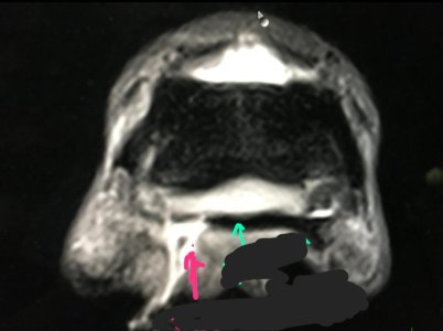
Number 1 MRI of a hoof but no indication of plane. The black area is probably fat depending on what imaging sequence was used but without anatomical landmarks the anatomy is very difficult. Not going to make a guess as to what the red or green arrows are pointing to as depending on MRI imaging sequence and 3D plane they could indicate many structures.I'll leave you with a few more feet to muse over whilst I go to bed
I have more pics of some of these from the initial problem to fixing it.
This is exactly what the vet showed me and explained on the screen on Lari's scan. Was significant damage to SDFT and DDFT.Skin
Superficial Digital Flexor Tendon (SDFT)
Deep Digital Flexor Tendon (DDFT)
Check Ligament
Suspensory Ligament
Number 1 MRI of a hoof but no indication of plane. The black area is probably fat depending on what imaging sequence was used but without anatomical landmarks the anatomy is very difficult. Not going to make a guess as to what the red or green arrows are pointing to as depending on MRI imaging sequence and 3D plane they could indicate many structures.
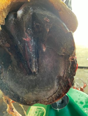
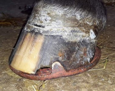
Number 2 Nasty under run sole abscess
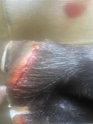
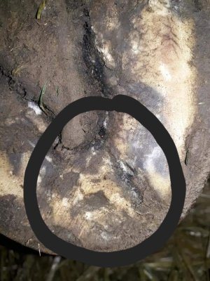
Number 3 Coronet band exit from a hoof abscess
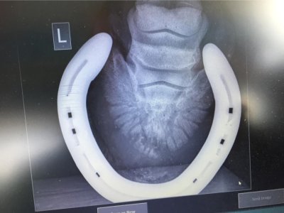
Number 4 Probably normal! The pedal bone when x-rayed from this aspect is always very "moth eaten" which is apparently normal. Shoes can still sometimes conceal delicate pathology. The area at the toe could be dirt but has no obvious pathology.
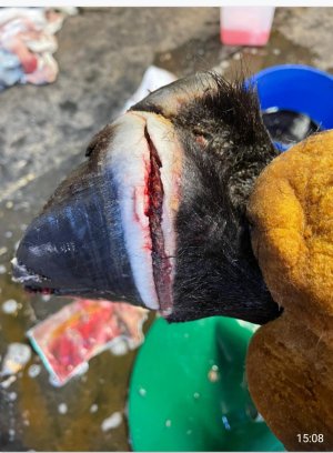
That is a curious angle - is it showing part of it magnified?
Fractured accessory carpal bone, back of the knee? (Probably not even a leg x-ray!I'll give you another bone one to play with.
 )
)